Coccidioides immitis
From IDWiki
Coccidioides immitis
- Dimorphic fungus in patients from arid areas in SW US and Mexico, which is mostly asymptomatic but can cause pulmonary disease, and rarely disseminated disease. Also called Joaquin Valley fever.
Background
Microbiology
- Dimorphic fungus with barrel-shaped arthroconidia in the septated hyphal filamentous form
- Coccidiomycosis also caused by Coccidioides posadasii outside of California
Epidemiology

- Found in thermic soils in arid regions
- May have an association with small desert mammals, though of unclear significance
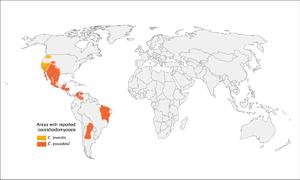
- C. immitis in California, C. posadasii outside of California (southwestern US, Mexico, Central and South America)
- Incidence in US is increasing over the past few decades, including emergence in Washington state
- Unlikely to be from increased detection
- 95% of cases are in California and Arizona
- Exposures include archaeological sites, desert exposures, dust storms, prisons
- It is a significant laboratory hazard
- There is a strain with a mutation that can cause aggressive invasive disease in younger adults
Pathophysiology
- Arthroconidia break off of the filamentous form and are inhaled (or inoculated)
- The then transform into invasive yeast form, characterized by spherules that contain hundreds of endospores
- The spherules rupture, releasing the endospores and continuing the cycle
Clinical Manifestations
- Variable, from asymptomatic or mild (60%), to symptomatic (40%), to disseminated and fulminant
- Incubation period of 1 to 3 weeks
- Common symptoms:
- General: fever, arthralgia, myalgia, night sweats
- Pulmonary: cough, chest pain
- May be a common cause of community-acquired pneumonia in some endemic regions.
- Non-specific maculopapular rash (days to weeks after initial fever)
- May have peripheral eosinophilia
- Pulmonary infection may present as an acute pneumonia to a chronic pneumonia with fibronodular or fibrocavitary appearance
- May also have a solitary nodule or a pleural effusion
- Skin infection can present at site of inoculation, or disseminated
- Can cause verrucous granulomatous lesions
- Erythema nodosum may be seen
- Erythema multiforme
- Non-specific examenthem
- Osteomyelitis and native joint septic arthritis, with the vertebral being to the most common site of infection
- May also cause arthralgias
- CNS infection with meningitis, presenting as a chronic headache weeks to months after initial infection
- Onset of meningitis is usually around 5 weeks following onset of respiratory symptoms
- Typically basilar meningitis but can be other
- Can cause prostatitis
- Symptoms may take weeks to many months to resolve
Desert rheumatism
- Erythema nodosum
- Fevers
- Arthralgias
Complications
- Chronic pulmonary disease:
- Residual lung nodules (~5%)
- Lung cavity (~5%)
- Extra-pulmonary dissemination (skin, bones, joints, CNS): ≤1%
Risk factors for disseminated disease
- Immunosuppression, including HIV/AIDS, pregnancy, TNF-α inhibitors, corticosteroids, and transplantation
- More common in non-Caucasian populations
- Age >65 years, diabetes, underlying cardiac or respiratory disease
- High complement fixation titre >1:16
Differential Diagnosis
- Infectious
- Bacterial: routine bacterial causes of pneumonia
- Fungal: Cryptococcus and other endemic fungi
- Mycobacterial: tuberculosis
- Viral?
- Non-infectious
Investigations
- CT chest1
- Acute disease: lobar and segmental consolidations (75%); multifocal consolidation; nodules; rarely, miliary disease or confluent nodules with cavitation; may have adenopathy or pleural effusions; may have ARDS
- Chronic changes: residual nodules, chronic cavities, persistent pneumonia with or without adenopathy, pleural effusion, and regressive changes
Diagnosis
- Direct microscopy: typical spherules
- Culture: fluffy white colonies on routine bacterial or fungal cultures, often within a few days (grows quickly)
- Serology
- EIA for IgM and IgG (used in Ontario)
- Sensitivity and specificity of 11-57% and 70-100% for IgM, and 53-69% and 95-99% for IgG, varies by test and laboratory2
- Immunodiffusion (ID) for IgM and IgG, plus complement fixation (CF) for IgG
- ID IgM on CSF for coccioidal meningitis
- Beta glucan in blood or CSF, but isn't specific
- Coccidioidal antigen (urine and serum), only useful in disseminated disease
- EIA for IgM and IgG (used in Ontario)
- For CNS involvement:
- Check opening pressure, send for fungal culture (25% sensitive), CSF serology (ID or CF, 30-60% sensitive), CSF coccidioidal antigen
- Glucose normal or low, protein high or normal, predominately lymphocytosis, may have eosinophils
- Rule out Cryptococcus and TB
Management
- Many do not need treatment, as infection is asymptomatic or self-limited
- Not indicated for asymptomatic pulmonary nodules or cavities in immunocompetent people
- Indications
- Significant, debilitating illness requiring hospitalization
- Extensive pulmonary involvement with diabetes, older age, or other comorbidities
- Chronic cavitary coccidioidal pneumonia
- Ruptured coccidioidal cavities
- Extrapulmonary soft tissue infections and bone and joint infections
- Meningitis
- High-risk populations: HIV with CD4<250, hematopoietic stem cell transplant, solid organ transplant
- For immunocompetent, ambulatory patients with acute pulmonary infection, one of
- Weight loss >10%
- Night sweats for 3 weeks
- Infiltrates on more than half of one lung or portions of both
- Prominent or persistent hilar adenopathy
- Anticoccidioidal complement fixation antibody titres over 1:16
- Inability to work
- Symptoms lasting >2 months
- Treatment regimens
- Fluconazole 400-800 mg/day
- May use itraconazole, voriconazole, posaconazole, or isavuconazole
- May use amphotericin B if life-threatening infections, pregnancy, or refractory to azole therapy
- Slight preference for itra for bone & joint infections
- Duration
- Acute pulmonary disease: 3 to 6 months, or longer
- Chronic pulmonary infection: 8 to 12 months, or longer
- Bone and joint disease: amphotericin B induction x 3 months followed by 3+ years of azoles
- Meningitis
- High-dose fluconazole until clinical improvement, then lifelong fluconazole 400 mg daily
- Daily therapeutic LP titrated to opening pressure (like cryptococcal meningitis)
Laboratory Exposure
- Evacuate lab, seal lab, call Biosafety Officer on call
- Assess risk to each individual exposed
- Lower early in culture when it's yeast-like, higher later when it's a well-grown filamentous fungus (usually by 7 to 10 days)
- Lifting the lid once likely less than breaking the container
- However, note that in lab exposures, the number of arthroconidia inhaled is often much higher than a natural inoculum, and attack rates are higher than natural exposure
- For exposed personnel
- Get baseline serology (if prior exposure, they're at lower risk)
- Treat with therapeutic-dose itra or fluc 400 mg daily for 6 weeks, as prophylaxis
- If pregnant, would have to consider prophylactic amphotericin; use of fluconazole may depend on trimester
- Repeat serology after prophylaxis; if seroconversion, then consider treating for another few months
- If they become symptomatic during or after prophylaxis, either with fever or respiratory symptoms, they should be further evaluated
- Serology can lag by 3 to 12 weeks following symptoms
- Continue to follow for up to 1 year
References
- ^ Cecilia M. Jude, Nita B. Nayak, Maitraya K. Patel, Monica Deshmukh, Poonam Batra. Pulmonary Coccidioidomycosis: Pictorial Review of Chest Radiographic and CT Findings. RadioGraphics. 2014;34(4):912-925. doi:10.1148/rg.344130134.
- ^ Ian H. McHardy, Bridget Barker, George R. Thompson. Romney M. Humphries. Review of Clinical and Laboratory Diagnostics for Coccidioidomycosis. Journal of Clinical Microbiology. 2023;61(5). doi:10.1128/jcm.01581-22.