Echinococcus granulosus
From IDWiki
Echinococcus granulosus
Background
Microbiology
- Cestode in the Echinococcus family
- Multiple strains cause hydatid cysts
- Strains have host specificity and are organised by genotype (G1 to G10):
- Echinococcus granulosus sensu stricto (G1 to G3), the most common cause worldwide
- Echinococcus equinus (G4), with no human cases reported
- Echinococcus ortleppi (G5)
- Echinococcus canadensis (G6 to G10)
Life Cycle
- Carnivores are the definitive host (canines, felids, or hyenids), where the protoscolex develops into a cestode (after 4 to 7 weeks) and releases eggs
- Eggs are infectious on release, and can be viable in the environment for months or longer
- Intermediate hosts are usually herbivors (sheep, goats, cattle, camels, and cervids), where the oncosphere hatches, penetrates the intestine, and migrates to the target organ, where it encysts to create the metacestode (or hydatid cyst)
- Within the cyst, protoscolices develop to form a germinal layer
- The protoscolices are ingested by the definitive host
Risk Factors
- Exposure to sheep or other livestock, especially when dogs are kept and fed offal
Clinical Manifestations
- Larval form can affect any organ, though most commonly the liver (80%) or lungs (20%)
- E. canadensis may be more likely to go to brain
- Cysts grow by 1 to 50 mm annually, and can spontaneously rupture
- Symptoms are caused by compression of nearby structures or by rupture
Diagnosis
- Based on clinical picture, imaging, and serology
Ultrasound

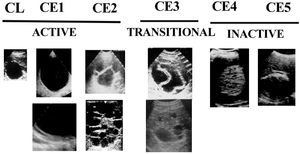
- Standardized ultrasound classifications have been created by the WHO1
- Active disease (CE1-CE2): cysts are developing and are usually fertile
- Transitional disease (CE3): cysts are starting to degenerate but the protoscoleces are still viable
- Inactive disease (CE4-CE5): cysts are fully degenerated and are now calcifying
- Pathognomonic features include:
- Unilocular anechoic lesion that are round or oval with a clear, well-defined wall, and souble line sign
- Multivesicular or multiseptated cysts with a wheel-like appearance
- Unilocular cysts with daughter cysts that may have a honeycomb-like appearance
- Cysts with floating laminated membranes with or without daughter cysts
References
- ^ WHO Informal Working Group. International classification of ultrasound images in cystic echinococcosis for application in clinical and field epidemiological settings. Acta Tropica. 2003;85(2):253-261. doi:10.1016/s0001-706x(02)00223-1.